
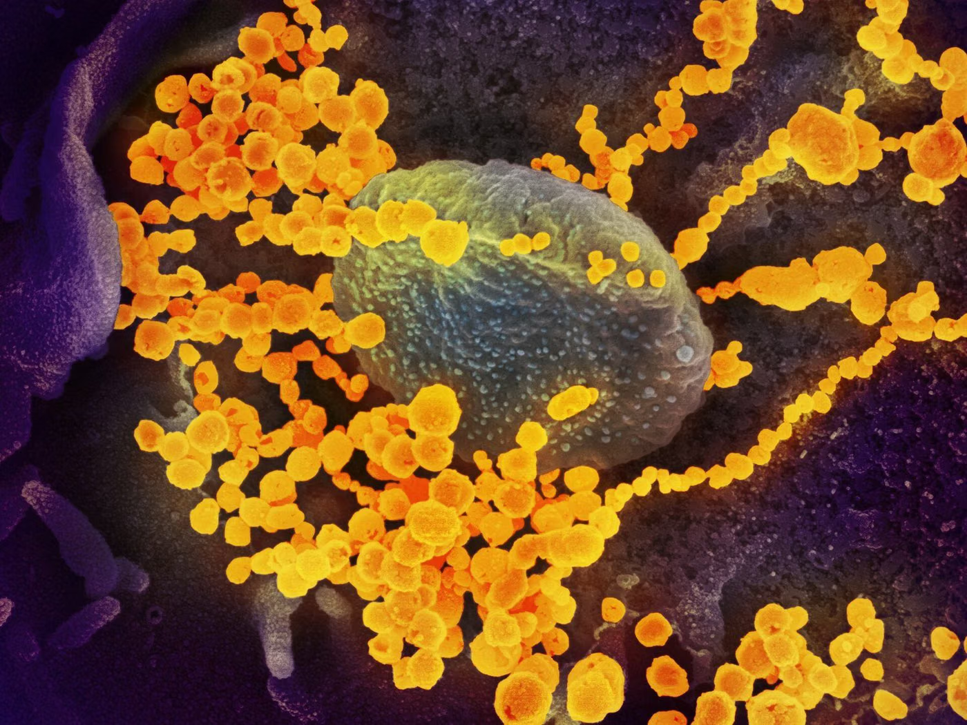
What Is SEM Analysis?
A scanning electron microscope scans a focused electron beam over the sample surface and collects the backscattered and secondary electrons from the surface. From these signals, a surface image with high depth of field is formed at very high magnifications.
Purpose of SEM Analysis
The purpose is to image surface details at a scale optical microscopes cannot resolve. Fracture-surface examination, coating cross-section measurement, weld structure, particle size and surface defect analysis are done with this technique.
Which Products Is SEM Analysis Applied To?
- Fractured or damaged metal parts
- Coating and thin-film cross-sections
- Powder and particulate materials
- Electronic components and solder joints
- Polymer and composite fracture surfaces
- Particles collected on a filter
In Which Sectors Is SEM Analysis Used?
Standards Related to SEM Analysis
- ISO 16700 (micromarking calibration — SEM magnification)
- ASTM E986 (SEM performance evaluation)
- ASTM E2809 (forensic SEM/EDS examinations)
SEM Analysis Conditions
- acceleration voltage
- It is selected according to the sample; Low voltage is preferred in insulators
- display mode
- Secondary electron (topography) or backscattered electron (composition contrast)
- Vacuum
- High vacuum; low vacuum mode for moist or insulating samples
- Kaplama
- Gold or carbon coating is applied to non-conductive samples
Equipment for SEM Analysis
- Scanning electron microscope
- Secondary and backscattered electron detectors
- Sputter coater
- Sample cutting, mounting and polishing infrastructure
- Image processing and measurement software
Sample Requirements for SEM Analysis
- The sample must fit within the vacuum chamber
- Non-conductive samples require a thin conductive coating; this process is permanent on the surface
- The sample is mounted and polished for cross-section examination — this is destructive
- The surface must not be cleaned for fracture-surface examination — it must be sent as-is
- Samples containing oil, moisture or volatiles disrupt the vacuum
Duration of SEM Analysis
An examination session is generally completed within hours. Cross-section preparation and examining many regions extend the duration to a few working days.
SEM Analysis Procedure
- 1The sample is cut and, if required, mounted and polished.
- 2A thin coating is applied if not conductive.
- 3The sample is placed in the vacuum chamber and vacuum is established.
- 4Accelerating voltage and detector are selected.
- 5The relevant regions are imaged at different magnifications.
- 6Measurements (coating thickness, particle size, etc.) are made with software.
- 7Findings are reported together with the images.
SEM Analysis Acceptance Criteria
The acceptance limit for surface defects or coating thickness comes from the product standard or specification; the microscope only measures.
Evaluation of Results
An SEM image is qualitative evidence; a numerical result is meaningful only if the magnification calibration is current and measurement regions are representatively selected. In fracture-surface interpretation, the fracture type (brittle, ductile, fatigue) is read from the morphology.
SEM Analysis Report
- Sample description and purpose of examination
- Sample preparation steps and coating information
- Instrument conditions (voltage, detector, magnification)
- Images showing the examined regions and a scale bar
- Measurements made and observations
- A statement that the results relate only to the regions examined
Accreditation
EUROLAB holds internationally recognised accreditation obtained from accreditation bodies party to the IAF and ILAC multilateral recognition arrangements, under ISO/IEC 17025 for laboratory testing, ISO/IEC 17020 for inspection and surveillance, and ISO/IEC 17021-1 and ISO/IEC 17065 for system and product certification. This chain ensures the reports and certificates issued are accepted worldwide.
Whether this test can be offered within accredited scope depends on your product and the standard applied; since accreditation is method-based, the scope is confirmed in writing at the quotation stage.
Scope and authorisationsRelated Regulations
- Failure analysis requirements in aviation and defence procurement specifications
- Surface characterisation in medical device technical files
Pages Where This Method Is Used
Analytical Methods — What Does This Group of Methods Do?
The section below describes the field that the SEM Analysis test belongs to; the conditions and acceptance criteria of this particular test are given in the sections above. The scope specific to your product is confirmed in writing at the quotation stage.
Analytical method pages are pages about a MEASUREMENT TECHNIQUE rather than about a test. The difference matters: "salt spray testing" is a test and answers one question; "ICP-MS" is a technique and is used in dozens of different tests. Sometimes the user is looking not for the test but for the technique — because the method name is what appears in their specification.
This distinction becomes decisive in choosing a laboratory. When a customer asks for "heavy metal analysis", the real question is by which technique it will be done: XRF is fast and non-destructive but does not distinguish compounds; AAS is economical for a single element; ICP-OES screens multiple elements; ICP-MS sees trace levels. The same sample gives results with different detection limits and different uncertainties across the four techniques.
Method selection is made against three criteria. DETECTION LIMIT: is the concentration sought within the range the method can see? MATRIX COMPATIBILITY: is the sample matrix (plastic, food, soil, metal) one the method can handle, and is pre-treatment required? SPECIFICATION LINK: does the product standard or legislation mandate a particular method? The third often overrides the first two — the specification binds the method.
Method pages build a CHAIN. A technique is linked to the test pages where it is used, to the standards those tests rest on, to the legislation those standards are tied to and ultimately to market access regimes. The example chain in the source document works like this: XRF → metal analysis → RoHS compliance → heavy metal restriction → coating pages. Whichever link the user enters from, they reach the others.
Sample preparation is the invisible half of the result in this family and is a greater source of error than the technique itself. The digestion method, degree of grinding, extraction solvent and dilution ratio change the result markedly. The same sample on the same instrument gives a different result with different preparation; the definition of the method therefore covers preparation too.
Calibration and verification are the third determining component. An instrument is calibrated with certified reference material, the calibration curve is verified periodically and a control sample is run in each batch. Without these steps, a number produced is not traceable even if the instrument is working correctly.
Method groups in this field
Elemental analysis
ICP-OES, ICP-MS, AAS and XRF. They differ in detection limit, matrix tolerance and multi-element capacity.
Chromatography
GC, GC-MS, HPLC, LC-MS/MS and ion chromatography. It separates a mixture into its components and measures each separately.
Spectroscopy
FTIR, Raman, UV-Vis and optical emission. For material identity, functional group identification and alloy composition.
Thermal analysis
DSC, TGA, DMA and dilatometry. Transition temperatures, composition, decomposition and dimensional behaviour.
Microscopy and imaging
Optical microscopy, SEM-EDS, radiography and tomography. Structure, defects and elemental distribution.
Classical (wet) chemistry
Titration, gravimetry and colorimetry. Used as a reference method and in confirming instrumental results.
Commonly confused in this field
- XRF is a screening tool; it sees elements and does not distinguish compounds. A decision on a restricted compound requires confirmation by wet chemistry.
- The phrase "not detected" is not proof of absence; it is read together with the detection limit of the method.
- The specification binds the method. A technically better method is not accepted if the specification calls for another.
- Sample preparation is part of the result; a report that does not state the preparation is not reproducible.
- Results obtained by different techniques cannot be compared directly; their detection limits and uncertainties differ.
- A method page is not a test page; which method applies to which test comes from the product standard.
Which decisions these results support
- Matching the method written in the specification to the laboratory's scope
- Method selection according to detection limit and matrix compatibility
- Confirming an instrumental result with a reference method
- Eliminating deviations arising from sample preparation
Related Tests
Frequently Asked Questions About SEM Analysis
Can elemental analysis be performed with SEM?
SEM alone gives an image. An EDS detector is needed for elemental information; this combination is described on a separate page as SEM-EDS.
Can the sample be recovered?
If a conductive coating has been applied or a cross-section taken, the sample cannot be restored to its original state. This is communicated before the analysis.